


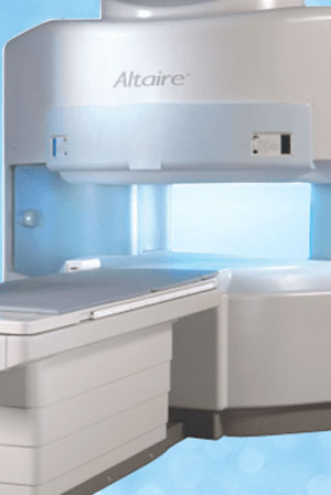
Open MRI
Our Open MRI
For your comfort, we have an open MRI for those who are claustrophobic and can accommodate pediatric patients and those up to 500 pounds. The Hitachi .7 Altaire is a powerful MRI capable of high resolution imaging in a comfortable setting to reduce patient anxiety.
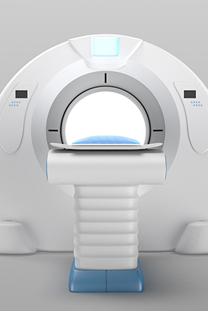
Computed Tomography
What is Computed Tomography
Computed Tomography (CT) sometimes called “CAT” scan, is a scanning method used to see inside the body, slice by slice, something standard x-ray exams are unable to do. The CT exam, a fusion of x-ray equipment and computer analysis, provides a highly detailed image of the internal anatomy, revealing the location, size and shape of abnormal growths. Computed tomography (CT) scanning is used to gather images on organs, blood vessels, brain, bones, and the spinal cord. In some cases, a contrast material or dye is injected during a scan to make structures more visible.
In the case of the multi-detection CT, remarkable three-dimensional pictures are made, further allowing physicians to diagnose certain diseases earlier and more precisely. Spiral CT scanners, using a multi-slice system, perform a continuous scan of larger areas in less than half the time of standard CT scanners. They also provide precise two and three-dimensional images that were previously only visible through more invasive procedures.
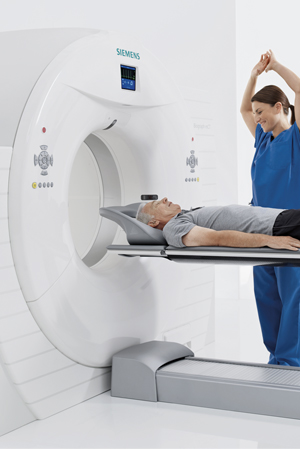
PET-CT Technology
Advanced Diagnostic Technology
The fastest, most accurate and comfortable PET-CT technology
- 128-slice, high-resolution imaging
- Great for claustrophobic and bariatric patients – widest and shortest tunnel combination in healthcare
- Large field of view for highest accuracy
CareFirst Imaging Center in Schererville is equipped with the Siemens Biograph mCT — an innovative positron emission tomography/computed tomography (PET-CT) scanner, which offers accurate, fast and comfortable exams for a wide range of patients.
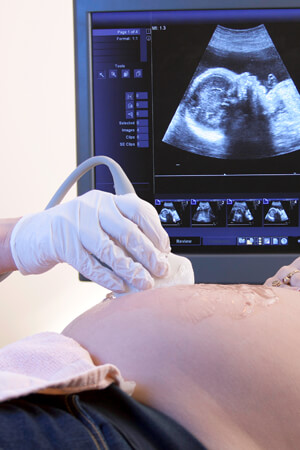
Ultrasound
What is Ultrasound
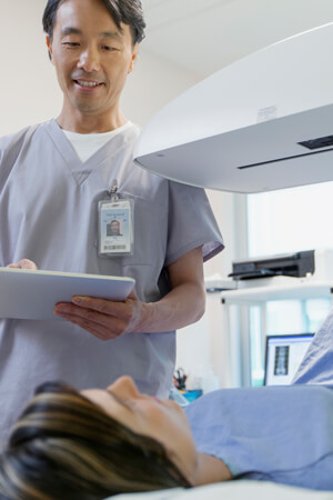
Ultrasound, also known as sonography, uses high-frequency sound waves to gather real-time images within the body. It is a safe and painless way of examining organs such as the heart, liver, kidneys and bladder, as well as showing movement of internal tissues and organs. Physicians can see blood flow and heart valve functions which can help to diagnose and monitor a variety of heart conditions. No ionizing radiation (x-ray) is used in ultrasound imaging. Ultrasound is a non-invasive medical test that helps physicians diagnose and treat medical conditions. Conventional ultrasound displays the images in thin, flat sections of the body.
A Doppler ultrasound study may be part of an ultrasound examination.
Bone Density
What is Bone Densitometry?
Bone Densitometry is a non-invasive means of measuring the density of bones. DEXA is widely used for the evaluation of osteoporosis. Bone density scanning measures the bone mineral density, which is related to bone mass. Decreased bone mass causes decreased bone strength and subsequent risk of fractures. Bone mass peaks in men and women at about age 30. Men are generally large and hence have a greater overall amount of bone mass. Both women and men experience a loss of bone mass of 3-5% per decade. Once women reach menopause, the rate of bone loss accelerates to 1% – 3% per year. This rate is slowed by hormone replacement.Fractures are the first clinical signs of osteoporosis. It is desirable to identify patients who have osteoporosis before development of fractures. People are considered at risk of developing osteoporosis based on certain risk factors and whether pre or post-menopausal.

3D Mammography
What is 3D Mammography?
Otherwise known as tomosynthesis, 3D Mammography enables our radiologists view breast tissue layer by layer, helping them to see inside the breast with previously unimaginable detail, especially in areas with overlapping tissue. 3D mammography is available for all women seeking screening mammograms, but it’s also recommended for women who have a family history of breast cancer, who had previous breast biopsies, and/or have dense breasts.
The 3D mammogram experience is very similar to that of a traditional mammogram. Like a traditional mammogram, the technologist will position you, compress your breast and take images from different angles. It takes just a few seconds longer for each image.
X-Ray
What is X-Ray
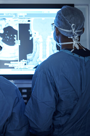
X-ray has been called one of the most significant advances in all of medical history. It is used in many different ways in medical diagnosis. An x-ray image is produced when a small amount of radiation passes through the body and strikes a sheet of sensitive film placed on the other side of the body. The ability of x-rays to penetrate tissues and bone varies according to the tissue composition and mass. Bone, which contains calcium, does not let much radiation through and results in white images on the x-ray film. The lungs, which are filled with air, allow nearly all x-rays to strike the film resulting in a black film image.
Methodist CareFirst Imaging Center
SCHERERVILLE
7860 Burr St.
Schererville, IN 46375
219-738-5930
Fax 219-864-2910
Open Mon. – Fri.: 7:30am – 7pm | Sat.: 10am – 5pm